Discovery of prostaglandin receptors promoting implantation

DP/EP4 agonist as an implantation promoter applied for infertility care
Summary
- Prostaglandins (PGs)*1 are a series of bioactive lipids produced by various organs that exert pathological effects such as fever and pain, but were also known to be involved in reproductive processes such as parturition.
- In the uterus, PGs produced during implantation was known to play roles in enlargement of the implantation site (IS) (decidualization)*2, but the PG receptors that transmit this effect were unknown.
- In this study, we found that PGD2, in addition to PGE2, is produced in the uterus during implantation and acts on EP4 and DP receptors, respectively, as a promoter of decidualization. Therefore, we have shown that activation of either DP or EP4 receptors during implantation can induce decidualization.
- PGs are likely to work similarly in humans, and enhancing PG function with DP/EP4 agonists could improve implantation problems that cause infertility.
Outline of the study
A research group led by Professor Yukihiko Sugimoto and Assistant Professor Tomoaki Inazumi at Faculty of Life Sciences, Kumamoto University, in collaboration with Professor Yasushi Hirota and Project Assistant Professor Shizu Aikawa at Graduate School of Medicine, University of Tokyo, and Professor Toru Takeo at Kumamoto University Center for Animal Resources and Development, has demonstrated for the first time in the world that prostaglandin (PG) D2, a bioactive lipid produced in the uterus promotes decidualization via its receptor DP, that the PGE2-EP4 receptor pathway also promotes decidualization in parallel with this pathway, and that simultaneous blockade of both pathways disrupts decidualization. Based on this achievement, it is expected that the DP/EP4 receptor agonist will be effective in treating infertility, especially in improving implantation defects on the uterine side.
The research results were published in the American scientific journal “Journal of Lipid Research” on Friday, August 30, 2024
Background
Implantation, the phenomenon of the embryo joining and infiltrating the uterus, is the starting point of pregnancy. It is only when the embryo and uterus interact with each other at the appropriate time that the embryo's growth is stimulated and its supporting tissues are formed. For example, when the embryo attaches and is taken into the uterus, this is the starting point for decidualization*2 and pregnancy. In fact, defects in decidualization can lead to infertility, but the detailed mechanism therein remained unclear. Therefore, from the viewpoint of infertility treatment, the earlier elucidation of the molecular mechanism of decidualization has been long awaited. It is known that COX-2, a PG-producing enzyme, plays a central role in implantation and that PG-like bioactive lipids produced by COX-2 promote implantation and decidualization, but the type of PG receptor that mediates these actions is unknown.
Research outline
The research group focused on the important role of cAMP signaling in stromal cells for uterine decidualization and analyzed the expression of PG receptors, DP, EP2, and EP4, all of which stimulate the cAMP signaling. The group found that DP receptor is expressed in the luminal epithelium during the implantation period together with COX-2, EP2 and EP4 receptors. Furthermore, DP receptor is induced in mesometrial stromal cells upon implantation, whereas EP4 receptor is expressed in the anti-mesometrial stromal cells (Fig. 1). Utilizing the fact that COX inhibition*3 impairs decidualization, the group examined the restoring effects of each receptor agonist and found that the decidualization failure was restored by DP or EP4 agonists, but not by EP2 agonist (Fig. 2). The group also generated EP2/DP double-deficient (EP2/DPKO) mice and examined their implantation receptivity, which showed no abnormalities. When the EP4 blocker was administered to the KO mice, decidualization was markedly inhibited (Fig. 3). These results strongly suggest that the PGD2-DP and PGE2-EP4 receptor pathways are activated after implantation in the uterus to promote decidualization (Fig. 4) and that both pathways compensate for each other's functions and contribute to the execution of a series of implantation processes.
Significance
This study provides a new scientific understanding of the molecular mechanism underlying implantation and decidualization, and elucidates how multiple PG receptors compensate for each other's functions to achieve the implantation processes in order to nurture the life of the offspring.
Application
PG receptors are likely to function similarly in humans, and enhancing PG function with DP/EP4 receptor agonist is expected to prevent and treat implantation failure, which is a problem in infertility treatment.
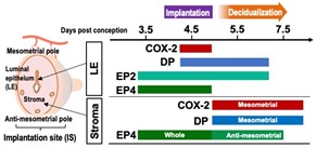
Fig. 1. Expression sites of PG receptors during peri-implantation period.
DP receptors were expressed in the luminal epithelium during the implantation period (Day 4.5 post-conception) along with COX-2, EP2 and EP4. At day 5.5, when decidualization undergoes, DP receptors are expressed adjacent to COX-2 in the mesometrial stromal cells, and EP4 receptors are expressed in the anti-mesometrial stromal cells.
DP receptors were expressed in the luminal epithelium during the implantation period (Day 4.5 post-conception) along with COX-2, EP2 and EP4. At day 5.5, when decidualization undergoes, DP receptors are expressed adjacent to COX-2 in the mesometrial stromal cells, and EP4 receptors are expressed in the anti-mesometrial stromal cells.
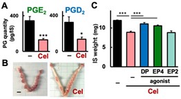
Fig. 2. COX-2 inhibitor-elicited decidualization failure was restored by DP or EP4 receptor agonists.
COX-2 inhibitor (Cel) administered at Day 4.5 decreased PGE2 and PGD2 levels in the implantation site (IS) (A) and weakened IS growth (B). (C) Cel-induced IS weight loss was restored by DP or EP4 agonists, but not by EP2 agonist.
COX-2 inhibitor (Cel) administered at Day 4.5 decreased PGE2 and PGD2 levels in the implantation site (IS) (A) and weakened IS growth (B). (C) Cel-induced IS weight loss was restored by DP or EP4 agonists, but not by EP2 agonist.
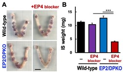
Fig. 3. EP4 blockade severely inhibits decidualization in EP2/DP-deficient (KO) mice.
EP4 blocker was administered to wild-type and EP2/DPKO mice at days 4.5 and 5.5 after transfer of wild-type embryos, and the uterus was examined at day 6.5. Only when EP4 blocker was administered to EP2/DPKO mice, IS growth was severely attenuated (A) and its weight was reduced to less than half (B).
EP4 blocker was administered to wild-type and EP2/DPKO mice at days 4.5 and 5.5 after transfer of wild-type embryos, and the uterus was examined at day 6.5. Only when EP4 blocker was administered to EP2/DPKO mice, IS growth was severely attenuated (A) and its weight was reduced to less than half (B).
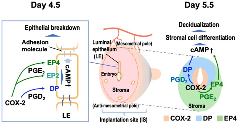
Fig. 4. Role of PGD2-DP and PGE2-EP4 pathways in decidualization.
(Day 4.5) DP, EP2, and EP4 receptors are expressed in the luminal epithelium and contribute to epithelial breakdown by PGD2 and PGE2 produced by COX-2. (Day 5.5) DP and EP4 receptors are expressed in the mesometrial and anti-mesometrial stroma, respectively, and promote decidualization by PGD2 and PGE2 produced by COX-2.
(Day 4.5) DP, EP2, and EP4 receptors are expressed in the luminal epithelium and contribute to epithelial breakdown by PGD2 and PGE2 produced by COX-2. (Day 5.5) DP and EP4 receptors are expressed in the mesometrial and anti-mesometrial stroma, respectively, and promote decidualization by PGD2 and PGE2 produced by COX-2.
Terminology
*1 Prostaglandins (PGs)
PGD2 and PGE2 are representative bioactive lipids synthesized by cyclooxygenase (COX) and specific synthases (Fig. 5). PGD2 exerts sleep induction and allergic responses, and PGE2 exerts fever, pain sensation, and inflammatory responses.
*1 Prostaglandins (PGs)
PGD2 and PGE2 are representative bioactive lipids synthesized by cyclooxygenase (COX) and specific synthases (Fig. 5). PGD2 exerts sleep induction and allergic responses, and PGE2 exerts fever, pain sensation, and inflammatory responses.
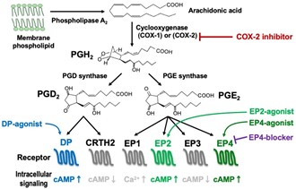
Fig. 5. Synthetic pathways and receptors of PGD2, PGE2.
When cells are exposed to various stimuli, arachidonic acid, a kind of fatty acid, is excised from cell membrane phospholipids by phospholipase A2. Arachidonic acid is converted to PG precursor (PGH2) by cyclooxygenase (COX-1 or COX-2), and further to PGD2 and PGE2 by PGD synthase and PGE synthase, respectively. The effects of PGD2 and PGE2 are exerted through two (DP and CRTH2) and four (EP1 to EP4) types of receptors, respectively, among which DP, EP2, and EP4 induce various cellular responses via the stimulation of intracellular cAMP production.
*2 Decidualization
Uterine tissue consists of the outer myometrium, the inner luminal epithelium, and stroma between them (Fig. 6). When the embryo attaches to the luminal epithelium and implantation occurs, the epithelium collapses and the embryo is infiltrated into the stroma. The peri-embryonic stromal cells then differentiate into decidual cells, proliferating to form the pre-placental tissue, which serves as a bed for the embryo. This phenomenon is called decidualization.
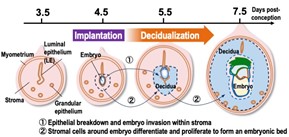
Fig. 6. Anatomy of the uterus and implantation/decidualization
*3 COX inhibitor
Aspirin is the most common Non-Steroidal Anti-Inflammatory Drugs (NSAIDs), which irreversibly inhibit COX (COX-1/2), the rate-limiting enzyme in PG production, thereby inhibiting PG biosynthesis. There are many other NSAIDs, including indomethacin, ibuprofen, and diclofenac. In this study, celecoxib (Fig. 7) was used to selectively inhibit COX-2 induced by implantation.
Aspirin is the most common Non-Steroidal Anti-Inflammatory Drugs (NSAIDs), which irreversibly inhibit COX (COX-1/2), the rate-limiting enzyme in PG production, thereby inhibiting PG biosynthesis. There are many other NSAIDs, including indomethacin, ibuprofen, and diclofenac. In this study, celecoxib (Fig. 7) was used to selectively inhibit COX-2 induced by implantation.
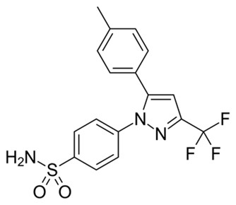
Fig. 7. Structure of celecoxib
Supporting information
This research was supported by a Grant-in-Aid for Scientific Research from the Japan Society for the Promotion of Science, Ono Medical Research Foundation, Naito Foundation, and Kanzawa Medical Research Foundation.
Paper information
Title: “Uterine prostaglandin DP receptor induced upon implantation contributes to decidualization together with EP4 receptor”
Authors: Risa Sakamoto, Takuji Fujiwara, Yuko Kawano, Shizu Aikawa, Tomoaki Inazumi, On Nakayama, Yukiko Kawasaki-Shirata, Miho Hashimoto-Iwasaki, Toshiko Sugimoto, Soken Tsuchiya, Satohiro Nakao, Toru Takeo, Yasushi Hirota, Yukihiko Sugimoto
Journal: Journal of Lipid Research
doi:10.1016/j.jlr.2024.100636
URL:https://www.jlr.org/article/S0022-2275(24)00141-X/fulltext
| Contact information Kumamoto University, Faculty of Life Sciences Professor Yukihiko Sugimoto Phone:096-371-4357 E-mail:ysugi@kumamoto-u.ac.jp |