The discovery brings hope for the development of new oral cancer treatments.
Extracellular vesicles derived from radioresistant oral cancer cells regulate radiation-induced apoptosis via their encapsulated microRNAs, and cause the target cancer cells to acquire radioresistance.
Researchers found that measuring microRNAs (miR-503-3p) in the blood of patients with oral cancer could predict the effects of radiotherapy and patient prognosis.
A new mechanism for the acquisition of radioresistance in oral cancer involving extracellular vesicles has been revealed which may lead to the development of new diagnostic and therapeutic methods.
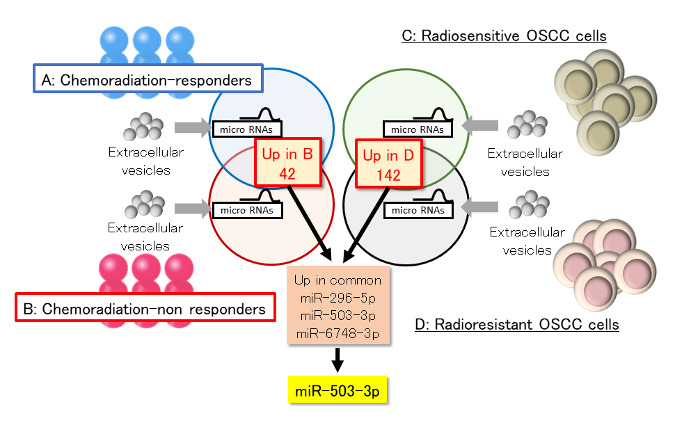
Identification of microRNA involved in radioresistance
(Left) The expression levels of microRNAs in extracellular vesicles extracted from chemoradiation-responder and chemoradiation-non responder patients were compared, and 42 microRNAs were picked up that were elevated in the inactive patients. (Right) The expression levels of microRNAs in extracellular vesicles extracted from radiation-sensitive and radiation-resistant cells were compared, and 142 microRNAs were picked up. (Mid-bottom) Preliminary experiments were conducted using three macroRNAs whose expression was up-regulated in both, and finally miR-503-3p was identified as a microRNA involved in radioresistance.
A research group led by Associate Professor Ryoji Yoshida and Professor Hideki Nakayama from the Department of Oral and Maxillofacial Surgery in Kumamoto University (Japan) has analyzed the extracellular vesicles (EVs) of radiation-resistant oral cancer cells and the microRNA contained within them, and discovered a new mechanism by which microRNA imparts radioresistance to surrounding radiation-sensitive oral cancer cells. The researchers believe that that their discovery may lead to the development of new diagnosis and treatment methods for radiation-resistant oral cancer.
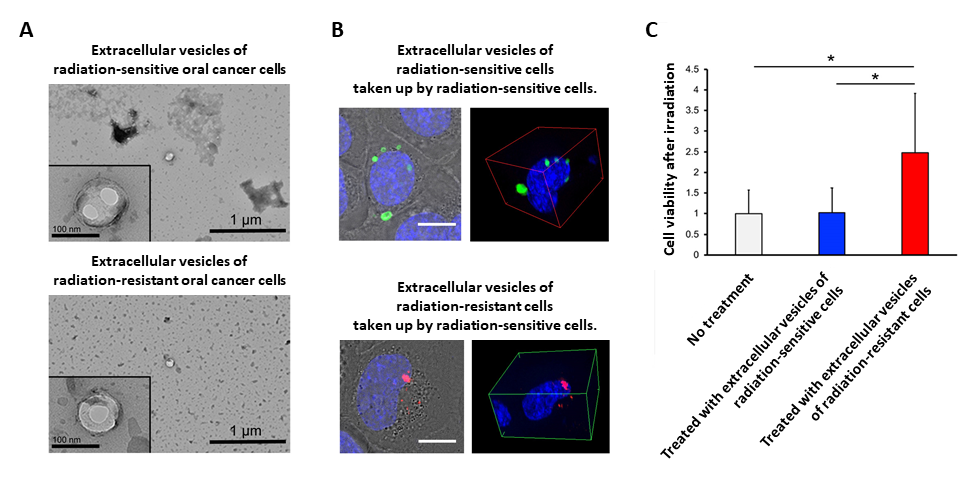
Effect of extracellular vesicles isolated from oral cancer cells on the radioresistance of oral cancer
A) Transmission electron microscopy images of extracellular vesicles isolated from radiation-sensitive (top) and radiation-resisitant (bottom) cells. The lower left square is a magnified image.
B) Fluorescence microscopy images of extracellular vesicles taken up by radiation-sensitive cells. Upper: Extracellular vesicles derived from radiation-sensitive cell (green) are incorporated into a radiation-sensitive cell (blue). Lower: Extracellular vesicles derived from a radiation-resistant cell (red) are incorporated into a radiation-sensitive cell (blue).
C) Change in radioresistance when extracellular vesicles are incorporated into a radiation-sensitive cell. The graphs show the cell viability under each condition. Gray: no treatment, Blue: treated with extracellular vesicles of radiation-sensitive cells, Red: treated with extracellular vesicles of radiation-resistant cells. From Figs. 1D, 2D & 2E of Yamana K., et.al., Journal of Extracellular Vesicles, 2021. The image has been modified for use and clarity within this release. This image is made available under CC BY-NC 4.0 and any further distribution must follow the terms of this license.
Radiation therapy is the second most used treatment, after surgery, for oral cancer. However, some oral cancers are resistant to radiation therapy and may recur or worsen. The prognosis for patients with radiation resistant oral cancer is poor.
Cancer cells are believed to acquire radiation treatment resistance by “talking to each other” using a form of cell-to-cell communication to share various information. The focus of most research has been on EVs, which are released outside the cell, as the cellular communications toolbox because they contain a large amount of information in the form of genes and proteins. Furthermore, little research has been done on the relationship between EVs and oral cancer radiotherapy resistance.
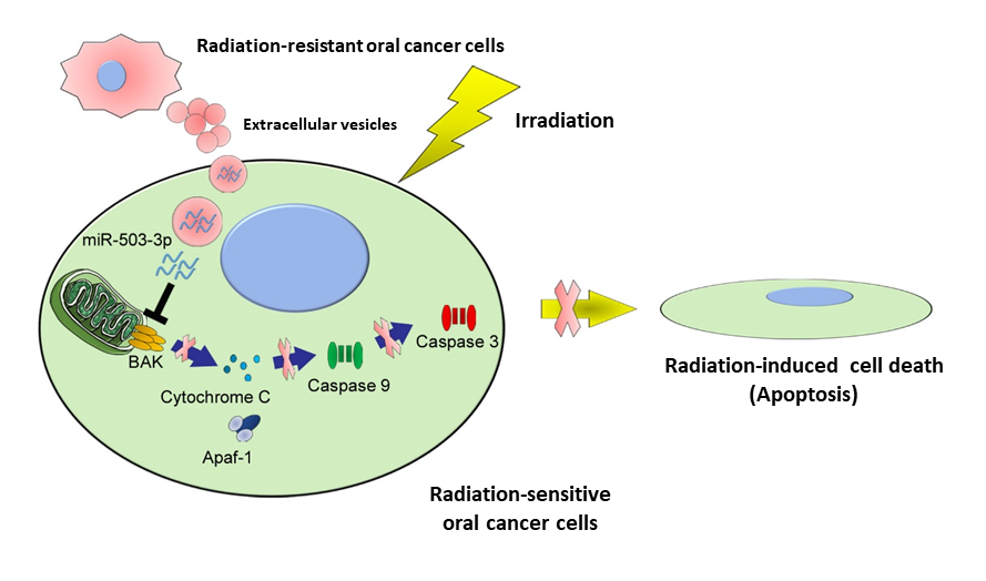
Mechanisms of radiation resistance acquisition in oral cancer cells
Extracellular vesicles released from radiation-resistant oral cancer cells are taken up by radiation-sensitive cells, which release miR-503-3p. The released miR-503-3p suppresses BAK, a protein that promotes apoptosis, thereby preventing radiation-induced cell death (apoptosis). As a result, radiation-sensitive cells acquire radioresistant. From Fig. 6E of Yamana K., et.al., Journal of Extracellular Vesicles, 2021. The image has been modified for use and clarity within this release. This image is made available under CC BY-NC 4.0 and any further distribution must follow the terms of this license.
In what they believe to be a world first, the Yoshida & Nakayama research group successfully isolated EVs released from radiation-resistant oral cancer cells. These EVs were then added to radiation-sensitive oral cancer cells which revealed that radioresistance was acquired by radiation-sensitive cancer cells through the transfer of microRNA, one of the pieces of information contained within the EV toolbox. The microRNA (miR-503-3p) is able to suppress apoptosis (cell death) after irradiation, thereby making the formally radiation-sensitive cells radioresistant. The researchers believe that this mechanism highly contributes to the establishment of radioresistant oral cancer within the body.
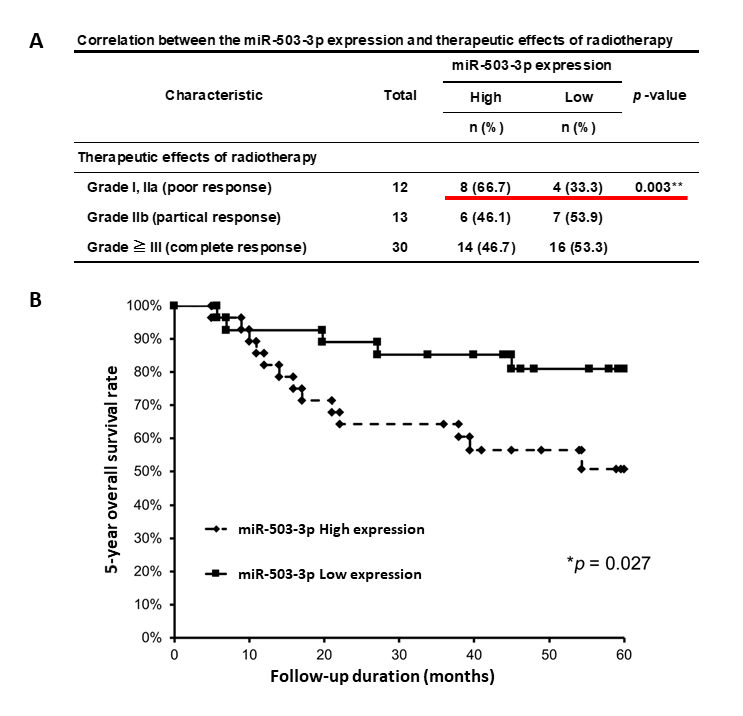
Additionally, oral cancer patients with high expression levels of the miR-503-3p microRNA in their blood had poorer treatment effects and a worse prognosis. In other words, the researchers found that the amount of microRNA in the blood may predict both the therapeutic effect and the prognosis of radiotherapy in oral cancer patients.
On uncovering this new mechanism for radiation resistance acquisition by oral cancer, Professor Nakayama said, “Our research may lead to the development of new diagnostic and therapeutic methods for this type of cancer. We hope to soon begin animal experiments with the goal of developing a therapeutic strategy that targets the EVs secreted by radioresistant oral cancer."
Relationship between the expression level of microRNAs (miR-503-3p) in blood and radiotherapy effect and patient prognosis
A) Relationship between the expression level of miR-503-3p and radiotherapy effect. Patients with high expression level of miR-503-3p have poor treatment effect (red underlined area).
B) Relationship between miR-503-3p expression level and patient prognosis. 5-year survival rate is significantly lower in patients with high miR-503-3p expression level (dashed line). From Table 2 & Fig. 7A of Yamana K., et.al., Journal of Extracellular Vesicles, 2021. The image has been modified for use and clarity within this release. This image is made available under CC BY-NC 4.0 and any further distribution must follow the terms of this license.
This research was published online on 11 December 2021 in the Journal of Extracellular Vesicles.
[Usage Restrictions]
Image credits belong to Dr. Ryoichi Yoshida unless otherwise indicated in the figure caption. To use any of the media contained within this release elsewhere, a reference to the original work (this release) should be included and other restrictions must be followed as indicated.